本文主要是介绍comsol 缝合_如何“缝合”视网膜,应该怎么做?,希望对大家解决编程问题提供一定的参考价值,需要的开发者们随着小编来一起学习吧!
comsol 缝合
And they recommend you to “sew it on”. And you doubt — and this is exactly what you need? And how safe is it? But nothing bothers — then why? Or maybe they want to earn money on me? And the first thing you start is to read posts on the Internet, what a “independent” expert like Google will say.
他们建议您“缝上它”。 您怀疑-这正是您所需要的吗? 它有多安全? 但是什么都没有打扰-那为什么呢? 也许他们想在我身上赚钱? 首先,您要阅读互联网上的帖子,像Google这样的“独立”专家会说些什么。
And in the future everything depends on your discipline and attention to your own health. You can get to the ophthalmologist-laser specialist, who will be the last resort, and he will do preventive laser coagulation.
将来,一切都取决于您的纪律和对自己健康的关注。 您可以找眼科医生激光专家,他们将是万不得已的方法,他将进行激光预防性凝结。
Or «forget it» for everything and continue to live as before — nothing bothers you. What is the risk?
或“忘了一切”,让一切继续像往常一样生活-没有什么困扰您。 有什么风险?

To understand what works and how, we need knowledge of the anatomy of the eye.
要了解什么有效以及如何起作用,我们需要了解眼睛的解剖结构。
The retina (retina) of the eye is the inner membrane, therefore, to inspect it, you need to look into the eye cavity through the pupil. The wider the pupil, the more the surface of the retina will be able to see the ophthalmologist. Therefore, to examine the fundus in the maximum amount, you need to bury the drops that can cause cycloplegia — a condition where the pupil is wide and does not respond to light. This reversible state, albeit a rather unpleasant one, passes in a couple of hours, but it makes it possible to look into the most “secret” zones of the retina.
眼睛的视网膜是内膜,因此,要检查它,您需要通过瞳Kong观察眼腔。 瞳Kong越宽,视网膜表面越能见到眼科医生。 因此,要检查最大量的眼底,您需要掩盖可能导致睫状肌麻痹的滴剂,这种情况是瞳Kong宽而对光没有React的情况。 这种可逆的状态虽然令人不快,但在几个小时内就过去了,但它可以观察到视网膜最“秘密”的区域。
The human retina consists of two parts.
人体视网膜由两部分组成。
The back is photosensitive; front end — not sensitive to light. Conventionally, the separation takes place «at the equator»: the department after the equator is the visual, or functionally active part of the retina, the neuronal retina, which we see. We don’t see a department around the equator, and problems arise there.
背面是光敏的; 前端-对光不敏感。 按照惯例,分离发生在“赤道”:赤道之后的部分是我们看到的视网膜的视觉或功能活跃部分,即神经元视网膜。 我们在赤道周围没有部门,那里出现了问题。
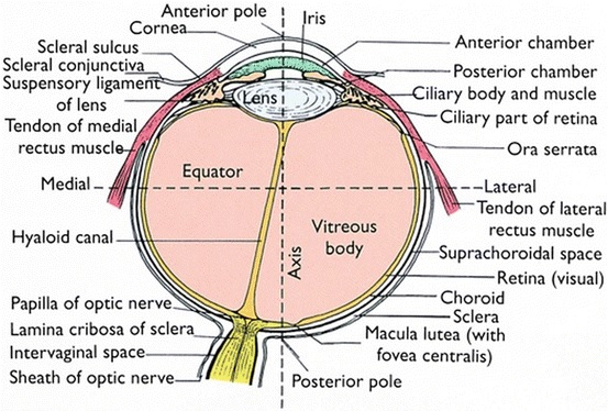
The visual part of the retina is its posterior, or photosensitive, section, a complex structure containing at least 15 types of neurons connected to each other by intercellular connections with well-developed microvilli and closure belts. These specialized compounds provide the difference in electrical potential due to the transport of ions between the surfaces. Through the intermediate layer, including conductor neurons, these cells send processes to the brain. These processes converge in the papilla of the optic nerve, forming the optic nerve.
视网膜的视觉部分是其后部或感光部分,是一个复杂的结构,包含至少15种神经元,这些神经元通过与发达的微绒毛和闭合带的细胞间连接相互连接。 这些专门的化合物由于离子在表面之间的传输而提供了电位差。 这些细胞通过包括导体神经元在内的中间层将过程发送至大脑。 这些过程会聚在视神经乳头中,形成视神经。
But since none of these types of outgrowths are anatomically connected with photoreceptors, these areas can easily be separated from each other, for example, when retinal detachment develops.
但是,由于这些类型的产物均未在解剖学上与感光体相连,因此,例如在视网膜脱离发展时,这些区域很容易彼此分离。
是否可以独立检测视网膜的周围萎缩? (Independently it is possible to detect the peripheral distrophy of the retina?)
If a person has a problem in the visual part, he has complaints of reduced vision, is plagued by all sorts of unpleasant symptoms such as flashes, distortions, etc.
如果一个人的视觉部分有问题,他会抱怨视力下降,并受到各种不适症状的困扰,例如闪光,变形等。
And if the problems are in the «blind» zone, then there will be no complaints. Peripheral retinal dystrophy — an invisible danger.
如果问题出在“盲区”,那就不会有任何投诉。 周围性视网膜营养不良-一种看不见的危险。
The peripheral zone of the retina is practically invisible with the usual standard examination of the fundus without pupil dilation and inspection with special lenses that increase the angle and area of the review. But it is precisely at the periphery of the retina that dystrophic (degenerative) processes often develop, which are dangerous because they can lead to tears and retinal detachment. Changes in the periphery of the fundus — peripheral dystrophies of the retina — can occur in both short-sighted and far-sighted people, and in persons with normal vision.
视网膜的外围区域实际上是通过常规的眼底标准检查而看不见的,而无需瞳Kong散大,并使用特殊的镜片检查以增加检查的角度和面积。 但是营养不良(变性)过程经常在视网膜的周围发展,这很危险,因为它们会导致眼泪和视网膜脱离。 近视眼和远视眼以及视力正常的人都可能发生眼底周围的改变(视网膜的周围营养不良)。
This common and serious human disease can be effectively treated using laser surgery.
可以使用激光手术有效地治疗这种常见且严重的人类疾病。
齿龈周围营养不良的可能原因 (Possible causes of dystrophy in grindle periphery)
The causes of peripheral dystrophic changes in the retina are not fully understood.
视网膜周围营养不良性改变的原因尚不完全清楚。
Dystrophy may occur at any age, with the same probability in men and women.
营养不良可在任何年龄发生,男女患病率相同。
There are many predisposing factors — this is myopia, regardless of the degree of minus, heredity, inflammatory diseases and eye injuries, head injuries and, of course, age.
有很多诱发因素-这是近视,与负重,遗传,炎症性疾病和眼外伤,头部受伤以及年龄的大小无关。
Common diseases also have an effect: hypertension, atherosclerosis, diabetes, past infections and intoxications.
常见疾病也有影响:高血压,动脉粥样硬化,糖尿病,既往感染和中毒。
Approximately it happens this way: on the periphery of the retina, for some reason, nutrition is deteriorating, the vessels empty, this leads to metabolic depletion and thinning of local functionally modified areas.
大概是这样发生的:由于某种原因,在视网膜的外围,营养恶化,血管排空,这导致新陈代谢耗竭和局部功能改变的区域变薄。
Under the action of physical exertion, falls, conditions associated with lifting to a height or underwater immersion, acceleration, jerks and weight transfer, vibration, dystrophic altered areas can cause ruptures.
在体力消耗,跌倒,抬高或在水下浸入,加速,猛拉和重量转移,振动,营养不良的改变区域相关的条件下,可能导致破裂。
However, it has been proven that in people with myopia, peripheral degenerative changes in the retina occur much more frequently, because with myopia, the length of the eye increases, resulting in stretching of its membranes and thinning of the retina in the periphery.
但是,已经证明,在近视患者中,视网膜周边变性的改变更频繁发生,因为在近视患者中,眼睛的长度增加,从而导致其膜的拉伸和周边视网膜的变薄。
Distrophy和有什么不一样 (What is the difference between distrophy)
The first is the size and localization. They can be multiple or single, small or huge, occupy a local zone or be located around the entire circumference of the fundus.
首先是规模和本地化。 它们可以是多个或单个,较小或巨大,占据局部区域或位于眼底的整个周围。
The second, important, is what structures of the eye are affected. Peripheral retinal dystrophy is divided into peripheral chorioretinal (PCRD), when only the retina and choroid are affected, and peripheral vitreorecorioretinal dystrophy (PWHT) — with involvement in the degenerative process of the vitreous body. What is the vitreous body and what is happening in it is described here: “Flying midges” and “glassy worms” in the eyes, or where do “broken pixels” in the vitreous body come from.
其次,重要的是眼睛的哪些结构受到影响。 当仅视网膜和脉络膜受到影响时,周围视网膜营养不良分为外周脉络膜视网膜营养不良(PCRD)和外周玻璃体视网膜视网膜营养不良(PWHT),这与玻璃体的退化过程有关。 玻璃体是什么,其中发生了什么:眼睛中的“飞虫”和“玻璃蠕虫”,或玻璃体中的“残破像素”来自何处。
The type of dystrophy without participation of the vitreous is more benign. Such options include, for example, dystrophy of the type “cobblestone pavement”.
没有玻璃体参与的营养不良类型更为良性。 这样的选择包括例如“鹅卵石路面”类型的营养不良。
If in the dystrophy zone there are adhesions with the vitreous body, then often tractions (cords, adhesions) form between the modified vitreous body and the retina. It is highly likely that for any deformity it will pull the retina behind itself into the eye. Such adhesions, joining one end to the thinned area of the retina, many times increase the risk of ruptures and subsequent detachment. And this is all the chances of losing sight or degrading the optical quality of the eye to a few percent of the norm.
如果在营养不良区与玻璃体发生粘连,那么在修饰的玻璃体和视网膜之间通常会形成牵引力(绳索,粘连)。 对于任何畸形,极有可能将其自身后面的视网膜拉入眼睛。 这种粘连将一端连接到视网膜变薄的区域,多次增加了破裂和随后脱落的风险。 这是所有失去视力或将眼睛的光学质量降低到正常水平的百分之几的机会。
Such tears and dystrophies occur imperceptibly for the patient and are found not only in the near-sighted, but also in the far-sighted, and in people with absolutely normal optics of the eye.
这样的眼泪和营养不良对于患者而言是不明显的,并且不仅在近视眼中而且在远视眼中以及在具有完全正常的眼睛视力的人中都发现。
结束时发生了什么? (What is happening with a close?)
In myopic people, as a rule, the axial length of the eye is more than 24 mm (the average parameter, the measurement of which tells us about the progression of myopia). There is a risk that the eye, increasing (and an increase in the eye — this is, in fact, myopia) begins to pull the vitreous body (because the rest of the tissues stretch very little). Dystrophic chorioretinal processes on the periphery of the retina (“lattice degeneration”, “snail track”, etc.) lead to thinning of the retina and its ruptures.
通常,在近视人群中,眼睛的眼轴长度超过24毫米(平均参数,其测量值可以告诉我们近视的发展)。 存在眼睛增大(和眼睛增大,实际上是近视)的风险开始拉动玻璃体的原因(因为其余组织的拉伸很小)。 视网膜外围的营养不良性脉络膜视网膜病变(“晶格变性”,“蜗牛痕迹”等)导致视网膜变薄及其破裂。
With them, one can live as a whole without any symptoms, but the risk of retinal detachment in these cases is very high.
有了它们,人们可以整体生活而没有任何症状,但是在这些情况下视网膜脱离的风险非常高。
诊断周围营养不良和可缩回间隙 (Diagnostics of peripheral dystrophy and retractable gaps)
Peripheral dystrophies are asymptomatic and dangerous. Sometimes patients come with complaints of floating cloud and midges before the eyes, less likely to be disturbed by «sparks» and «lightning» at the periphery. That is, the symptoms are rare and scanty.
周围营养不良是无症状和危险的。 有时,患者会抱怨眼前浮起云朵和蚊虫,因此不太可能被周围的“火花”和“闪电”所干扰。 即,症状很少且很少。
And then it all depends on the ophthalmologist — will he persuade you to expand the pupil and see how the retina should be? Can you see problems and interprets what you see correctly? Whether it is enough to equip an office for diagnostics — it will simply not be possible to see with an ophthalmoscope flashlight, you need special lenses (contact or contactless). Is there a necessary diode laser in the clinic or will it be necessary to run and search for a clinic and a specialist who will strengthen the retina?
然后,这完全取决于眼科医生-他会说服您扩大瞳Kong,看看视网膜应该如何吗? 您能看到问题并正确解释您所看到的吗? 无论是否足以为诊断设备配备办公室-检眼镜手电筒根本无法看到,您需要特殊的镜片(接触式或非接触式)。 诊所中是否有必要的二极管激光器?是否有必要运营和寻找可以增强视网膜的诊所和专科医生?
Full diagnostics of peripheral dystrophy and “silent” tears (without retinal detachment) is possible when examining the fundus in conditions of maximum medical expansion of the pupil using a special lens, which allows you to view the most “remote” areas of the retina to the equator. If necessary, you can even use sclerocompression — indentation of the sclera as if inside the eyeball, thus the retina is shifted from the periphery to the center, with the result that some peripheral areas inaccessible for inspection become visible. This is all done after instillation of a local anesthetic for superficial freezing of the eye. The procedure will worsen your near vision somewhat, photophobia will increase by a couple of hours, but all these symptoms always pass on average in a couple of hours. So do not refuse it at all!
当使用特殊的镜片在瞳Kong最大程度地扩大瞳Kong的情况下检查眼底时,可以对周围营养不良和“无声的”眼泪(无视网膜脱离)进行全面诊断,这使您可以查看视网膜最远的区域。赤道。 如有必要,您甚至可以使用巩膜压迫术-像在眼球内部那样对巩膜进行压痕,从而使视网膜从周围转移到中心,从而使一些难以检查的周围区域变得可见。 这些都是在滴注局部麻醉剂以使眼睛浅表冻结之后完成的。 该过程会稍微恶化您的近视力,畏光将增加几个小时,但所有这些症状通常平均会在几个小时内消失。 所以根本不要拒绝它!
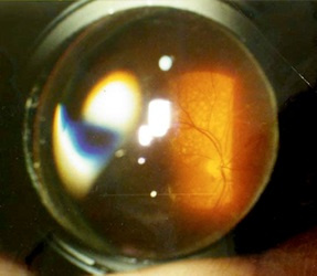
By the way, for inspection, you need to use not a flashlight, as is done in most clinics, but special lenses for examining the fundus. There is a universal contact lens — Goldman's lens. This flat lens with a system of three mirrors is very widely used in the examination and laser coagulation of the anterior segment of the eye and retina.
顺便说一句,进行检查时,您不需要像大多数诊所那样使用手电筒,而是需要使用特殊的镜片检查眼底。 有一个通用隐形眼镜-高盛镜头。 这种带有三面镜系统的平面透镜非常广泛地用于检查眼和视网膜的前段并进行激光凝结。
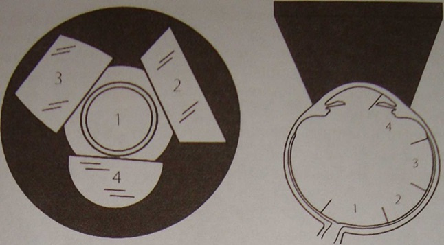
In each mirror, we see different parts of the retina, and, turning it clockwise, we get complete information. Something like this:
在每个镜子中,我们都可以看到视网膜的不同部分,顺时针旋转它可以获取完整的信息。 像这样:
And this is the scheme in which mirror which department of the eye we inspect:
这是我们检查哪个眼部的方案:
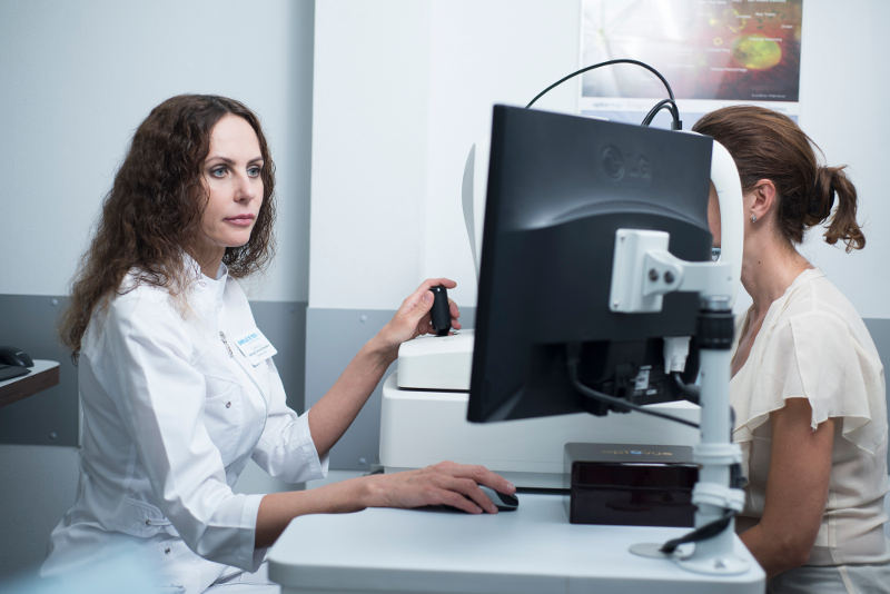
To date, there are also special digital cameras with which you can get a color image of the periphery of the retina in the presence of dystrophy and rupture zones.
迄今为止,还存在一些特殊的数码相机,在存在营养不良和破裂区域的情况下,您可以使用它们获得视网膜周围的彩色图像。
Well, and most importantly — the ophthalmologist must not only pretend that he is looking at the lens, he must be able to see and interpret what he has seen. My experience in treating detachments and problems with the vitreous body and retina indicates a sad situation.
好吧,最重要的是,眼科医生不仅必须假装自己正在看镜片,还必须能够看到并解释所见。 我在治疗玻璃体和视网膜的脱离和问题方面的经验表明情况很糟糕。
In half of the clinics sit doctors who do not even dilate the pupil for examination. Half of the remaining half looks with a direct ophthalmoscope — it shines a flashlight into the eye, while at best it sees only the center. The next half of the remaining quarter has lenses and looks, but does not understand anything in what they see!
在一半的诊所中坐着医生,他们甚至不扩大瞳Kong进行检查。 其余一半的一半用直接检眼镜看—它使手电筒射入眼睛,而充其量只能看到中心。 其余四分之一的下半部分都有镜头和外观,但看不到任何东西!
So, only 10-15% of patients receive high-quality diagnostics! The remaining 85-90% of doctors rely on luck, or refer to the fact that they do not have the necessary equipment.
因此,只有10-15%的患者会接受高质量的诊断! 其余的85-90%的医生依靠运气,或者指的是他们没有必要的设备。
The main necessary «equipment» for the diagnosis — the doctor's eyes and his professional intelligence.
诊断所需的主要“设备”是医生的眼睛和专业知识。
激光凝集周围萎缩和折返区 (Lasercoagulation of peripheral distrophy and zinary return zones)
The source of radiation in coagulation lasers is a diode-pumped solid-state laser.
凝结激光器中的辐射源是二极管泵浦的固态激光器。
The goal of this treatment is to prevent retinal detachment.
这种治疗的目的是防止视网膜脱离。
Perform preventive barrier coagulation of the retina in the field of dystrophic changes or border laser coagulation around an existing gap. An impact on the retina along the edge of a dystrophic focus or rupture is performed, resulting in a “gluing” of the retina to the underlying eye shells at the points of exposure to laser radiation. Let me remind you that we are talking about those areas of the retina with which we «do not see», therefore after this procedure and the narrowing of the pupil, the vision is completely restored to the original.
在营养不良变化领域或在现有间隙周围进行边界激光凝结时,对视网膜进行预防性屏障凝结。 沿着营养不良性焦点或破裂的边缘对视网膜产生冲击,导致视网膜在暴露于激光辐射的点处“粘合”到下面的眼球。 让我提醒您,我们正在谈论的是我们“看不见”的视网膜区域,因此,在此过程和瞳Kong变窄之后,视力已完全恢复到原始状态。
The procedure is performed with a Goldman contact lens on the eye. This prevents involuntary eye movements and allows you to accurately focus the laser beam on the problem area.
该过程在眼睛上戴高盛隐形眼镜进行。 这样可以防止眼睛不随意移动,并使您可以将激光束准确聚焦在问题区域上。
The patient senses the laser action as flashes of bright light. As a rule, they do not cause any discomfort, but sometimes there may be a slight tingling, dizziness, or even nausea. The operation takes place in a sitting position. The eye itself is securely fixed, and the beam on a healthy retina is excluded.
病人感觉到激光在闪烁着明亮的闪光。 通常,它们不会引起任何不适,但有时可能会出现轻微的刺痛,头晕甚至恶心。 该操作在坐姿下进行。 眼睛本身固定牢固,并且排除了健康视网膜上的光束。
Laser coagulation is performed on an outpatient basis and is well tolerated by patients. It is necessary to take into account that the process of formation of adhesions takes some time, therefore, after laser coagulation is carried out, it is recommended to avoid large physical exertion, such as parachute jumping, weight lifting, etc. within 10-14 days. Otherwise, you can lead a normal life and drip 3-4 times a day preventive drops.
激光凝结是在门诊患者基础上进行的,并且患者可以很好地耐受。 有必要考虑到粘连的形成过程会花费一些时间,因此,在进行激光凝结之后,建议避免在10到14岁的时间内进行较大的体力劳动,例如跳伞,举重等。天。 否则,您可以过正常生活,每天滴滴3-4次预防性滴剂。

怀孕和分娩怎么样 (How about pregnancy and childbirth)
During pregnancy, it is necessary to inspect the fundus of the eye on the wide pupil at least twice — at the beginning and at the end of pregnancy. After delivery, an ophthalmologist examination is also recommended.
在怀孕期间,有必要至少在怀孕的开始和结束时两次检查宽大瞳Kong的眼底。 分娩后,还建议进行眼科医生检查。
If there is a problem, then in terms of up to 36 weeks we are quite calmly performing laser coagulation of the retina. Although, the sooner it is found, the better. Since in the last months to sit during this procedure is not very convenient. After 36-37 weeks, we believe that it is too late to coagulate — spikes may simply not have time to form.
如果有问题,那么在长达36周的时间内,我们将非常冷静地对视网膜进行激光凝结。 虽然,发现得越早越好。 由于在过去的几个月里坐这个程序不是很方便。 在36-37周后,我们认为凝结为时已晚-峰值可能根本没有时间形成。
Delimited zones of dystrophy, adhesions and tears are not a contraindication to natural childbirth, of course, if there are no obstetric contraindications.
当然,如果没有产科禁忌症,营养不良,粘连和泪液的划定区域也不是自然分娩的禁忌症。
Prevention of the dystrophic processes themselves on the periphery of the retina is possible in representatives of the risk group — these are myopic, patients with hereditary predisposition, children born after a severe course of pregnancy and childbirth, patients with hypertension, diabetes, vasculitis and other diseases for which deterioration of peripheral circulation is observed. Such people are also recommended regular preventive examinations by an ophthalmologist with an examination of the fundus.
风险人群的代表可以预防视网膜周围的营养不良过程,包括近视眼,遗传易感患者,严重妊娠和分娩后出生的孩子,高血压,糖尿病,血管炎和其他观察到外周循环恶化的疾病。 眼科医生还建议对这些人进行定期的预防检查,并检查眼底。
零售的周边分布和激光视线校正 (Peripheral distrophies of the retail and laser view correction)
In itself, the presence of dystrophy at the periphery is not a contraindication for laser vision correction. Preventive laser “stitching” is done on medical advances, regardless of whether there will be a subsequent correction or not.
就其本身而言,周围营养不良的存在并不是激光视力矫正的禁忌症。 预防性激光“缝合”是根据医学进展而进行的,而不管是否会进行后续矫正。
For LASIK, the retina is pre-strengthened, since the vacuum ring significantly increases the pressure, increasing the risk of retinal detachment.
对于LASIK,由于真空环会显着增加压力,因此会预先加固视网膜,从而增加视网膜脱离的风险。
For methods such as SMILE or FemtoLASIK, it is not so important when preventive peripheral laser coagulation of the retina (PPLK) is performed — before or after correction, as there is no increase in intraocular pressure.
对于SMILE或FemtoLASIK之类的方法,在进行预防性视网膜周边激光凝结(PPLK)时(在矫正之前或之后)并不重要,因为眼内压不会增加。
我个人的提示 (Tips from me personally)
- Never refuse to check the fundus with the expansion of the pupil for your own safety. Most often, retinal dystrophies are found by chance. 为了您自己的安全,切勿拒绝检查瞳Kong扩大时的眼底。 大多数情况下,偶然发现视网膜营养不良。
- If there are complaints about the occurrence of lightning, flashes, the sudden appearance of more or less number of floating flies go immediately to the reception of an ophthalmologist, this may indicate already existing retinal breakdown. 如果有人抱怨闪电,闪光灯,或多或少的浮蝇突然出现,请立即就医,这可能表示视网膜已经存在破裂。
- Do not refuse to examine the periphery with the help of the Goldman contact lens — this will allow you to see the most extreme areas of the retina. 不要拒绝在高盛隐形眼镜的帮助下检查周边-这将使您看到视网膜的最末端区域。
- You can fix the presence of problem areas on special digital equipment and evaluate them in relation to the entire fundus area. Serious clinics have this capability and equipment. 您可以修复特殊数字设备上存在的问题区域,并针对整个眼底区域进行评估。 严肃的诊所拥有这种能力和设备。
- When peripheral dystrophy and retinal tears are detected, laser treatment is performed to prevent retinal detachment, so the sooner you do this procedure, the better. 当检测到周围神经营养不良和视网膜裂Kong时,将进行激光治疗以防止视网膜脱离,因此越早执行此过程越好。
- The main way to prevent these complications is the timely diagnosis of peripheral retinal dystrophy in patients at risk, followed by regular monitoring and, if necessary, preventive laser coagulation. 预防这些并发症的主要方法是对有风险的患者及时诊断周围性视网膜营养不良,然后进行定期监测,必要时进行预防性激光凝结。
- Patients with existing pathology of the retina and patients at risk should undergo an examination 1 — 2 times a year. 存在视网膜病理的患者和有风险的患者应每年进行1-2次检查。
- Do not deprive the opportunity of your child to be born naturally — do timely preventive examinations during pregnancy and laser coagulation according to indications. 不要剥夺孩子自然出生的机会-怀孕期间应及时进行预防性检查,并根据适应症进行激光凝固检查。
翻译自: https://habr.com/en/company/klinika_shilovoy/blog/511142/
comsol 缝合
这篇关于comsol 缝合_如何“缝合”视网膜,应该怎么做?的文章就介绍到这儿,希望我们推荐的文章对编程师们有所帮助!

